2頭の犬における微胞子虫性角膜症
Microsporidial keratopathy in two dogs
Scurrell E, Manning S, Malho P, et al. Vet Ophthalmol. November 2019. doi:10.1111/vop.12726. PMID: 31758652
原文(PubMed)はこちら
微胞子虫性角膜症が2頭の犬で報告されている。両犬とも、多病巣性癒合性混濁を特徴とする片側性実質性角膜症を呈し、角膜切除標本の組織病理学的検査で診断が下された。1頭の犬でホルマリン固定パラフィン包埋角膜組織の透過型電子顕微鏡(TEM)を実施したところ、形態学的特徴はNosema種感染と一致していた。いずれの犬も最初に診断され、表層角膜切除術によって治療された。1頭の犬に抗真菌薬を追加投与し、2年後に局所再発後に全層角膜形成術を施行した。ミクロスポリジウム感染に起因する他の全身性病変は臨床的に同定されなかった。臨床的および診断的病理所見、治療、フォローアップについて論じている。
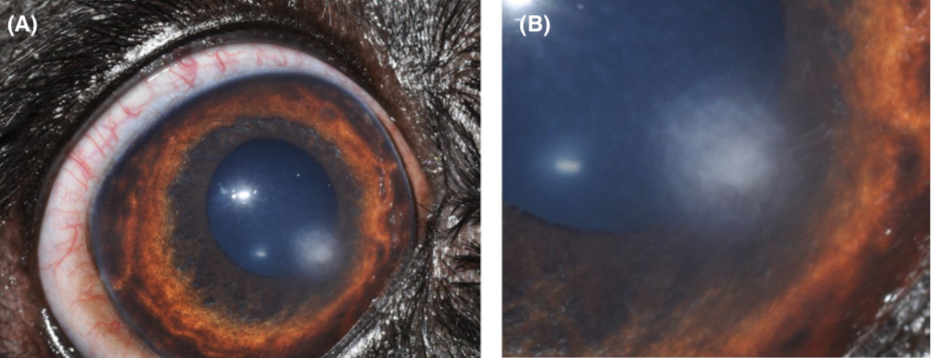
(症例2の前眼部写真。Figure 5より引用)
原文アブストラクト
A microsporidial keratopathy is described in two dogs. Both dogs presented with a unilateral stromal keratopathy characterized by multifocal coalescing opacities, and the diagnosis was made on histopathologic examination of keratectomy specimens. Transmission electron microscopy (TEM) on formalin-fixed, paraffin-embedded corneal tissue was performed in one dog, and the morphologic features were consistent with Nosema species infection. Both dogs were initially diagnosed and treated by superficial keratectomy. One dog received additional antifungal medication and underwent a penetrating keratoplasty following local recurrence two years later. No other systemic lesions attributable to the microsporidial infection were identified clinically. The clinical and diagnostic pathology findings, treatment, and follow-up are discussed.
