
光断層干渉計と蛍光眼底血管造影を用いた犬の突発性後天性網膜変性症の網膜の形態評価
Evaluation of retinal morphology of canine sudden acquired retinal degeneration syndrome using optical coherence tomography and fluorescein angiography.
光断層干渉計と蛍光眼底血管造影を用いた犬の突発性後天性網膜変性症の網膜の形態評価
Osinchuk SC, Leis ML, Salpeter EM, Sandmeyer LS, Grahn BH. Evaluation of retinal morphology of canine sudden acquired retinal degeneration syndrome using optical coherence tomography and fluorescein angiography. Vet Ophthalmol. 2018;19(4):319. doi:10.1111/vop.12602 / PMID:30136357
論文アブストラクト(PubMed)はこちら
PURPOSE:
To describe the optical coherence tomography (OCT) and fluorescein angiography changes in dogs with sudden acquired retinal degeneration syndrome (SARDS).
METHODS:
Retinal OCT was performed on 10 SARDS dogs and eight control dogs. Tomograms were collected in four quadrants around the optic nerve. Measurements were collected from the photoreceptor layer, the outer nuclear layer, the outer retina, the inner retina and the whole retina thickness in all quadrants. Sodium fluorescein was injected intravenously and serial fundic photographs were collected for a 5 minute period post-injection.
RESULTS:
In all quadrants, the outer nuclear layer (dorsal temporal P = 0.0000, dorsal nasal P = 0.0001, ventral temporal P = 0.0002, ventral nasal P = 0.000) and outer retina (dorsal temporal P = 0.0001, dorsal nasal P = 0.0002, ventral temporal P = 0.0054, ventral nasal P = 0.0084) measurements were significantly decreased in SARDS dogs. The whole retina thickness was significantly decreased in the dorsal temporal (P = 0.0082) and ventral temporal (P = 0.0428) retina. There were no significant differences in the photoreceptor layer thickness or inner retinal thickness between SARDS and control dogs. All SARDS dogs had a loss of definition of all of the photoreceptor bands on OCT. Two SARDS dogs had multifocal small retinal detachments and one of these dogs exhibited fluorescein leaking at the detachment sites.
CONCLUSIONS:
The significant reduction in the outer nuclear layer and the loss of band signals in the photoreceptor layers in dogs with SARDS identified on OCT support the previous histopathology findings. Small detachments may occasionally be detected on OCT and they may leak fluorescein.
目的:
突発性網膜変性症候群(SARDS)の犬の光干渉断層計(OCT: Optical coherence tomography)およびフルオレセイン血管造影の変化を報告すること。
方法:
10頭のSARDSの犬およびあ8頭の対照コントロールの犬において網膜のOCTを行った。OCT画像を視神経周囲の4象限で収集した。全ての象限において、光受容体層、外顆粒層、外側網膜、内側網膜及び網膜全体の厚さを測定した。フルオレセインナトリウムを静脈内注射し、注射後5分間、一連の眼底写真を記録した。
結果:
すべての象限において、外顆粒層(耳背側部 P = 0.0000、鼻背側 P = 0.0001、耳腹側P = 0.0002、鼻腹側 P = 0.000)および外側網膜(耳背側部P = 0.0001、鼻背側P = 0.0002、耳腹側 P = 0.0054、鼻腹側 P = 0.0084)の測定値は、SARDSの犬において有意に減少した。網膜全体の厚さは、外側背部(P = 0.0082)および外側腹部(P = 0.0428)網膜で有意に減少した。 SARDSと対照犬の間の光受容体層の厚さまたは網膜の厚さに有意差はなかった。すべてのSARDSの犬は、OCT上の光受容体バンドの全ての境界を失っていた。 2頭のSARDSの犬には多焦点の網膜剥離があり、これらの犬の1頭は剥離部位でフルオレセイン漏出を示した。
結論:
OCTで同定されたSARDSに罹患した外顆粒層の有意な減少および犬の光受容体層におけるバンドシグナルの消失は、以前の組織病理学的所見を支持した所見である。 OCTでは小さな剥離が検出されることがあり、フルオレセインが漏出することがある。
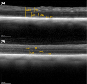
OCTでの正常犬とSARDSに罹患した犬の網膜の構造比較。(A) 正常な犬の網膜構造。(B)SARDSに罹患した犬の網膜の構造。Abbreviations: WR: whole retina, OR: outer retina, ONL: outer nuclear layer, PR: photoreceptor layer. (Fig 2より引用)