
白内障手術後の糖尿病犬における眼神経障害の回顧的解析
Retrospective analysis of ocular neuropathies in diabetic dogs following cataract surgery.
白内障手術後の糖尿病犬における眼神経障害の回顧的解析
Foote BC, Michau TM, Welihozkiy A, Stine JM. Retrospective analysis of ocular neuropathies in diabetic dogs following cataract surgery. Vet Ophthalmol 2018;165(Suppl 1):240. / PMID: 30095212
論文アブストラクト(PubMed)はこちら
To describe and compare the cumulative incidence and clinical progression of ocular neuropathies in diabetic dogs vs nondiabetic dogs following cataract surgery.
METHODS:
Medical records of 196 diabetic and 442 nondiabetic dogs who underwent cataract surgery between 2004 and 2015 were reviewed. The percentage of patients affected by neuropathy and potential risk factors were compared between groups.
RESULTS:
Patients with diabetes mellitus (DM) were 20.4 times more likely to develop an ocular neuropathy than patients without DM (12.24% vs 0.68%). Twenty-four diabetic patients were affected by mononeuropathies or polyneuropathies including Horner’s syndrome (n = 20), facial neuropathy (n = 5), and neurogenic keratoconjunctivitis sicca (NKCS) (n = 5). The odds of a diabetic patient developing Horner’s syndrome and NKCS were 86.3 and 20.7 times higher than a nondiabetic patient, respectively. The average duration of DM prior to diagnosis of neuropathy was 659 days (range 110-2390 days; median 559 days). Complete resolution was achieved in 10 of 22 neuropathies (45%) within an average of 248 days (range 21-638 days; median 187 days) after diagnosis.
CONCLUSIONS:
The odds of developing an ocular neuropathy, specifically Horner’s syndrome and NKCS, are statistically higher in diabetic patients compared to nondiabetic patients. Neuropathies were observed as a long-term complication in this group of diabetic patients, and complete resolution of the neuropathy was observed in less than half of the affected population.
KEYWORDS:
Horner’s syndrome; diabetes mellitus; diabetic neuropathy; facial neuropathy; neurogenic keratoconjunctivitis sicca; phacoemulsification
白内障手術後の糖尿病犬と非糖尿病犬の眼神経障害の累積発生率および臨床的な進行度を記述し比較すること。
方法:
2004年から2015年の間に白内障手術を受けた196頭の糖尿病犬、および442頭の非糖尿病犬の医療記録をレビューした。神経障害および潜在的な危険因子に冒された動物の割合をグループ間で比較した。
結果:
糖尿病(DM:diabetes mellitus)の動物は、DMでない動物よりも眼神経障害を発症する可能性が20.4倍高かった(12.24% vs 0.68%)。 24頭の糖尿病の動物は、ホーナー症候群(n = 20)、顔面神経障害(n = 5)、および神経性乾性角結膜炎(NKCS:neurogenic keratoconjunctivitis sicca )(n = 5)を含む、単神経障害または多発神経障害に罹患していた。ホーナー症候群およびNKCSを発症している糖尿病の動物のオッズは、非糖尿病の動物よりもそれぞれ86.3倍および20.7倍高かった。神経障害と診断される前のDMの平均期間は659日であった(範囲110-2390日;中央値559日)。診断後平均248日以内(21〜638日の範囲;中央値187日)に22症例中10症例(45%)の神経症が完全に回復した。
結論:
眼神経障害、特にホーナー症候群およびNKCSを発症する可能性は、非糖尿病の動物と比較して糖尿病の動物において統計学的に高い。この研究内の糖尿病の動物群では、神経障害が長期の合併症として観察され、神経障害の完全な回復が罹患した動物の半分未満で観察された。
キーワード:
ホーナー症候群、糖尿病、糖尿病性ニューロパチー、顔面神経障害、神経性乾性角結膜炎、水晶体超音波乳化吸引術
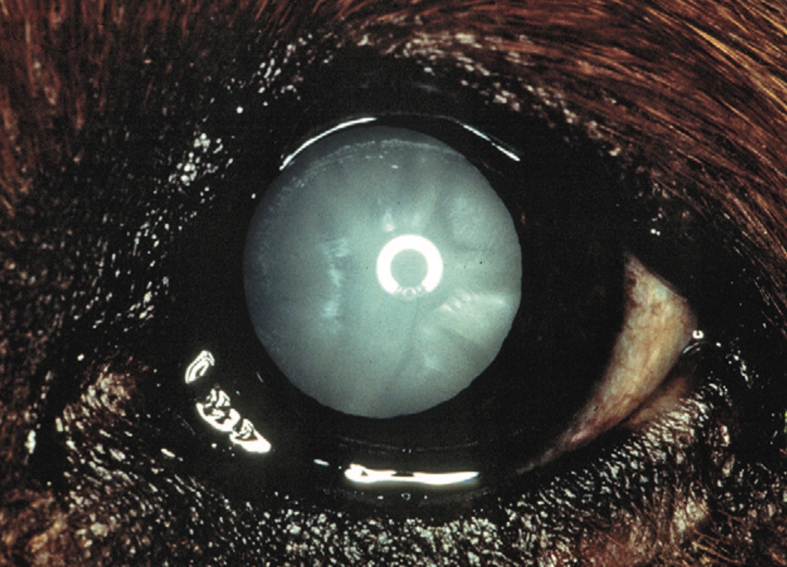
犬の糖尿病性白内障。 (Maggs DJ, Miller PE, Ofri R (eds): Slatter’s fundamentals of veterinary ophthalmology 5th ed. Fig 18-19 より引用)