
間葉系幹細胞で乾性角結膜炎を治療した犬の炎症マーカーCD4、IL-1およびIL-6の低下
Reduction in the inflammatory markers CD4, IL-1, IL-6 and TNFα in dogs with keratoconjunctivitis sicca treated topically with mesenchymal stem cells
間葉系幹細胞で乾性角結膜炎症を治療した犬の炎症マーカーCD4、IL-1およびIL-6の低下
Sgrignoli MR, Silva DA, Nascimento FF, et al. Reduction in the inflammatory markers CD4, IL-1, IL-6 and TNFα in dogs with keratoconjunctivitis sicca treated topically with mesenchymal stem cells. Stem Cell Res. 2019;39:101525. doi:10.1016/j.scr.2019.101525 / PMID:31430719
論文アブストラクト(PubMed)はこちら
論文アブストラクト
原文自動翻訳
Keratoconjunctivitis sicca (KCS) is of predominantly immune-mediated origin. Dogs are an excellent model for understanding this disease, as the origin of KCS in dogs is like that in humans. The objective of this study was to localize and quantify immunological markers, such as CD4 lymphocytes, interleukin (IL)-1, IL-6 and tumor necrosis factor alpha (TNFα), before and after topical treatment with mesenchymal stem cells (MSCs). Twenty-two dogs positive for KCS were topically treated with 50 μL (1 × 10⁶ MSCs) in the conjunctival sac and were evaluated for 6 months. The levels of the markers CD4, IL-6, IL-1 and TNFα were analyzed in conjunctival biopsy and cytology of the third eyelid gland by immunohistochemistry and immunocytochemistry. The results showed that before treatment, there was marked expression of all the markers (CD4, IL-6, IL-1 and TNFα), and after 6 months, there were significant (p < .05) reductions in the expression levels of all the markers. These results demonstrated that topical MSC treatment promotes a significant decrease in the expression levels of these inflammatory markers and could be used as adjuvant therapy in the treatment of KCS in dogs and humans. In addition, these markers can be excellent tools for diagnosing and analyzing the progression of KCS.
乾性角結膜炎(KCS)は主に免疫介在性の原因によるものである。 犬は、犬のKCSの原因がヒトのものと同様のものであるため、この疾患を理解するための優れたモデルである。 この研究の目的は、間葉系幹細胞(MSC)を用いた局所治療前後のCD4リンパ球、インターロイキン(IL)-1、IL-6および腫瘍壊死因子アルファ(TNFα)などの免疫学的マーカーの局在化および定量化することであった。 KCSの22頭のイヌで結膜嚢内に50 μL(1 × 10⁶ MSCs)を局所的に治療し,6 ヵ月間評価した。 マーカーCD4、IL-6、IL-1およびTNFαのレベルを、結膜生検および第3眼瞼腺の細胞診を免疫組織化学および免疫細胞化学によって分析した。 結果は、治療前には、すべてのマーカー(CD4、IL-6、IL-1およびTNFα)の顕著な発現が存在し、6ヶ月後にすべてのマーカーの発現レベルにおいて有意な低下(p < 0.05)が有意であることを示した。 これらの結果から、局所MSC治療は、これらの炎症性マーカーの発現レベルの有意な低下を促進し、イヌおよびヒトにおけるKCSの治療におけるアジュバント療法として使用することができることが実証された。 さらに、これらのマーカーは、KCSの進行を診断および分析するための優れたツールになる可能である。
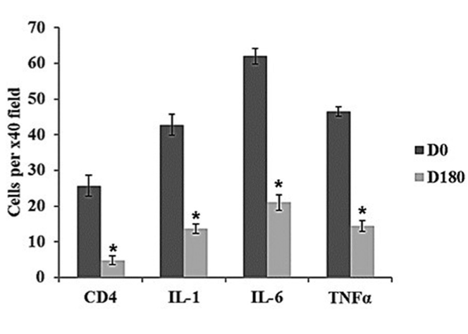
結膜でのCD4および各種サイトカイン陽性の細胞数。治療前と180日後で比較。 (Figure 3より引用)