
ボストンテリアにおける角膜内皮ジストロフィーの生体内in vivoイメージング:Fuchs角膜内皮ジストロフィーの自然発症イヌモデル
In Vivo Imaging of Corneal Endothelial Dystrophy in Boston Terriers: A Spontaneous, Canine Model for Fuchs’ Endothelial Corneal Dystrophy.
ボストンテリアにおける角膜内皮ジストロフィーの生体内in vivoイメージング:Fuchs角膜内皮ジストロフィーの自然発症イヌモデル
Thomasy SM et al. In Vivo Imaging of Corneal Endothelial Dystrophy in Boston Terriers: A Spontaneous, Canine Model for Fuchs’ Endothelial Corneal Dystrophy. Invest. Ophthalmol. Vis. Sci. 2016;57(9):OCT495–503. / PMID: 27454658
論文アブストラクト(PubMed)はこちら
PURPOSE:
Boston Terriers (BTs) have a greater prevalence of corneal endothelial dystrophy (CED), in comparison to other canine breeds. Similar to Fuchs’ endothelial corneal dystrophy (FECD), this condition is characterized by endothelial cell degeneration with secondary corneal edema. This study assessed corneal morphology using in vivo confocal microscopy (IVCM) and Fourier-domain optical coherence tomography (FD-OCT) in BTs with and without CED.
METHODS:
The corneas of 16 BTs with CED and 15 unaffected, age-matched BTs underwent clinical evaluation and were imaged using IVCM and FD-OCT. A two-sample t-test or Mann-Whitney rank sum test were used to statistically compare parameters between groups. Data are presented as mean ± SD or median (range).
RESULTS:
Mean age did not significantly differ between affected and unaffected dogs at 10.0 ± 2.0 and 10.6 ± 2.4 years, respectively (P = 0.437). Females (69%) were overrepresented among the CED-affected dogs. In CED patients, IVCM demonstrated endothelial polymegathism and pleomorphism. Corneal endothelial density was significantly less (P < 0.001) in dogs with CED (1026 ± 260 cells/mm2) versus age-matched controls (2297 ± 372 cells/mm2). Fourier-domain OCT demonstrated a significant increase (P < 0.01) in central corneal and endothelium-Descemet’s complex thickness in dogs with CED versus age-matched controls at 1019 (485-1550) or 536 (464-650) μm and 32 (22-56) or 25 (15-34) μm, respectively.
CONCLUSIONS:
Corneal endothelial dystrophy in BTs is a bilateral, adult-onset condition that shares many similarities with FECD. Thus, CED could serve as a spontaneous disease model to study the pathogenesis of and develop novel treatments for FECD.
ボストンテリア(BT:Boston Terriers)は、他の犬種と比較して、角膜内皮ジストロフィー(CED:corneal endothelial dystrophy)の有病率が高い。フックス角膜内皮ジストロフィー(FECD:Fuchs’ endothelial corneal dystrophy)と同様に、CEDは二次性の角膜浮腫を伴う内皮細胞変性を特徴としている。本研究では、CEDのBTと、CEDでないBTに対してin vivo共焦点顕微鏡(IVCM:in vivo confocal microscopy)とフーリエドメイン光干渉断層計(FD-OCT:Fourier-domain optical coherence tomography)を使用して角膜の形態を評価した。
方法:
CEDに罹患したBTの16角膜と同年齢のCEDでないBTの15角膜を臨床的に評価し、IVCMとFD-OCTを用いて画像を撮影した。 2標本t検定またはMann-Whitney順位和検定を使用して、グループ間のパラメータを統計的に比較した。データは平均値±SDまたは中央値(範囲)として表された。
結果:
平均年齢は、それぞれ10.0±2.0歳および10.6±2.4歳で、罹患犬と非罹患犬の間で有意差はなかった(P = 0.437)。メス(69%)は、CED罹患犬の間で有意に多かった。 CED患者では、IVCMで内皮細胞のサイズは非均一で、多形性を示した。角膜内皮密度は、年齢を一致させた対照(2297±372細胞/mm2)に対してCED(1026±260細胞/ mm2)を有するイヌにおいて有意に少なかった(P<0.001)。フーリエドメインOCTでは、角膜中央部はCED罹患BTとCED非罹患BTで、それぞれ1019(485-1550)と536(464-650)μmであり、内皮 – デスメ複合体の厚さはそれぞれ、32(22-56)と25(15-34)μmであり、CED非罹患BTと比較して、CED罹患BTで有意に増加した(P <0.01)。
結論:
BTにおける角膜内皮ジストロフィーは、FECDと多くの類似点を共有する、両側性の成犬で発症する疾病である。したがって、CEDはFECDの病因を研究し、FECDの新規治療法を開発するための自然発生疾患モデルとして役立つ可能性がある。
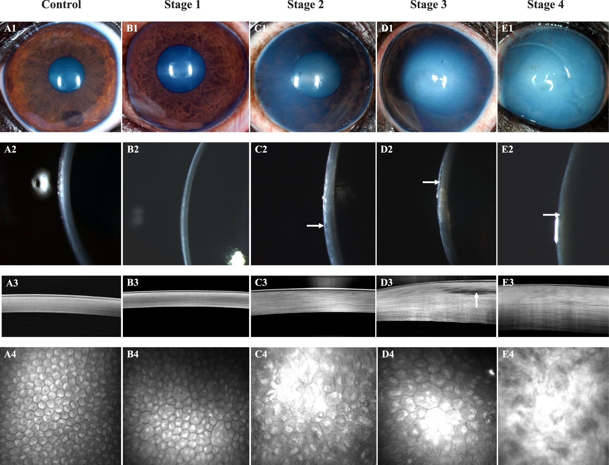
様々なステージのCED罹患ボストンテリアと非罹患ボストンテリアのスリットランプ、FD-OCT、IVCMの比較(FIg.1 より)