
生体内先端角膜イメージングと組織病理学を用いたジャーマン・ショートヘアーとワイアーヘアード・ポインターの角膜内皮ジストロフィーの表現型の特徴付け
Phenotypic Characterization of Corneal Endothelial Dystrophy in German Shorthaired and Wirehaired Pointers Using In Vivo Advanced Corneal Imaging and Histopathology.
生体内先端角膜イメージングと組織病理学を用いたジャーマン・ショートヘアーとワイアーヘアード・ポインターの角膜内皮ジストロフィーの表現型の特徴付け
Shull OR, Reilly CM, Davis LB, Murphy CJ, Thomasy SM. Phenotypic Characterization of Corneal Endothelial Dystrophy in German Shorthaired and Wirehaired Pointers Using In Vivo Advanced Corneal Imaging and Histopathology. Cornea 2018;37(1):88–94. / PMID: 29077583
論文アブストラクト(PubMed)はこちら
To evaluate corneal morphology using ultrasonic pachymetry (USP), Fourier-domain optical coherence tomography (FD-OCT), and in vivo confocal microscopy (IVCM) in 2 related canine breeds-German shorthaired pointers (GSHPs) and German wirehaired pointers (GWHPs)-with and without corneal endothelial dystrophy (CED). This condition is characterized by premature endothelial cell degeneration leading to concomitant corneal edema and is similar to Fuchs endothelial corneal dystrophy.
METHODS:
Corneas of 10 CED-affected (4 GSHP and 6 GWHP) and 19 unaffected, age-matched (15 GSHP and 4 GWHP) dogs were examined using USP, FD-OCT, and IVCM. A 2-sample t test or Mann-Whitney rank-sum test was used to statistically compare parameters between both groups. Data are presented as mean ± SD or median (range).
RESULTS:
Central corneal thickness determined using USP was significantly greater in CED-affected than in unaffected dogs at 1179 (953-1959) and 646 (497-737) μm, respectively (P < 0.001). Central epithelial thickness was found to be significantly decreased in CED-affected versus unaffected dogs at 47 ± 7.1 and 55 ± 7.1 μm, respectively (P = 0.011), using FD-OCT. With IVCM, corneal endothelial density was significantly less (P < 0.001) in 5 dogs with CED versus 19 unaffected controls at 499 ± 315 versus 1805 ± 298 cells/mm, respectively. CED-affected dogs exhibited endothelial pleomorphism and polymegethism, whereas CED-unaffected dogs had regular hexagonal arrangement of cells.
CONCLUSIONS:
GSHPs and GWHPs with CED exhibit marked differences in corneal morphology when compared with age-matched control dogs. These 2 CED-affected breeds represent spontaneous, large animal models for human Fuchs endothelial corneal dystrophy.
超音波パキメトリー(USP:ultrasonic pachymetry)、フーリエドメイン光干渉断層計(FD-OCT)、in vivo共焦点顕微鏡法(IVCM:in vivo confocal microscopy)を使用して、角膜内皮ジストロフィー(CED:corneal endothelial dystrophy)に罹患している、またはしていない、2つの関連犬種 – ジャーマン・ショートヘアード・ポインター(GSHP:breeds-German shorthaired pointers)およびジャーマン・ワイアーヘアード・ポインター(GWHP:German wirehaired pointers)を評価すること。これらの病態は、早期の内皮細胞変性を特徴とし付随する角膜浮腫をもたらす。そして人のフックス角膜内皮ジストロフィーと類似している。
方法:
10頭のCED罹患犬(4頭のGSHPおよび6頭のGWHP)および19頭の罹患していない同年齢の犬(15頭のGSHPおよび4頭のGWHP)の角膜をUSP、FD-OCT、およびIVCMを用いて調べた。 2標本t検定またはMann-Whitney順位和検定を使用して、両方のグループ間でパラメータを統計的に比較した。データは平均値±SDまたは中央値(範囲)として表された。
結果:
USPを用いて測定した中心角膜厚は、CED罹患犬と非罹患犬で、それぞれ1179(953-1959)および646(497-737)μmで、罹患していない犬よりもCED罹患犬において有意に大きかった(P <0.001)。 FD-OCTを用いて測定した中心上皮の厚さは、CED罹患犬と非罹患犬でそれぞれ47±7.1と55±7.1μmで、CED罹患犬で有意に減少した(P = 0.011)。 IVCMでは、角膜内皮密度は、CEDを有する5頭の犬において、499±315対1805±298細胞/ mmで、それぞれ19頭の非罹患対照とひかくして有意に低かった(P <0.001)。 CED罹患犬は内皮多形性および多細胞増殖を示したが、CED非罹患犬は規則的な六角形の細胞配列を示した。
結論:
CEDを有するGWHPおよびGWHPは、年齢の近い対照の犬と比較した場合、角膜の形態において著しい相違を示す。これらの2つのCED罹患品種は、人のフックス角膜内皮ジストロフィーの自然発生的な大型動物モデルとして適していることを表している。
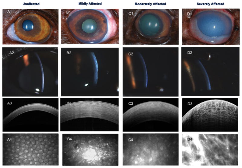
CEDに罹患した犬と非罹患犬のスリットランプ、FD-OCT、IVCMの比較(FIg.2 より)