緑内障を伴う猫の血管新生硝子体網膜症と前眼部異形成
Feline Neovascular Vitreoretinopathy and Anterior Segment Dysgenesis With Concurrent Glaucoma in Domestic Cats.
緑内障を伴う猫の血管新生硝子体網膜症と前眼部異形成
Beckwith-Cohen B, Hoffman A, McLellan GJ, Dubielzig RR. Feline Neovascular Vitreoretinopathy and Anterior Segment Dysgenesis With Concurrent Glaucoma in Domestic Cats. Veterinary Pathology 2018;:300985818798087. / PMID: 30222091
論文アブストラクト(PubMed)はこちら
論文アブストラクト
原文翻訳
Feline neovascular vitreoretinopathy (FNV) is a newly recognized rare condition affecting kittens and young domestic cats. This study investigated the clinical and pathologic findings in 22 cats with FNV. In affected cats, ophthalmoscopy of the fundus (when visible) revealed avascular peripheral retinae and epiretinal vascular membranes. Frequent nonspecific clinical findings were buphthalmos ( n = 21), medically uncontrollable glaucoma ( n = 22), and lenticular abnormalities ( n = 13). Anterior segment dysgenesis (ASD) was detected clinically in affected cats ( n = 6). The fellow eye was affected in 11 of 18 cats to a variable degree or appeared clinically normal in 7 of 18 cats. The globes were examined histologically and using immunohistochemistry for vimentin, glial fibrillary acidic protein (GFAP), synaptophysin, neurofilament, laminin, factor VIII-related antigen (FVIII-RA), and smooth muscle actin (SMA). Histologically, diagnostic features included laminin-positive epiretinal vascular membranes affecting the central retina, with an avascular peripheral retina and gliosis. Enucleated globes exhibited multiple additional abnormalities, including corneal disease ( n = 15), anterior segment dysgenesis ( n = 21), lymphoplasmacytic anterior uveitis ( n = 19), peripheral anterior synechiae ( n = 20), retinal degeneration ( n = 22), and retinal detachment ( n = 19). Gliotic retinae labeled strongly for GFAP and vimentin with reduced expression of synaptophysin and neurofilament, consistent with degeneration or lack of differentiation. While an avascular peripheral retina and epiretinal fibrovascular membranes are also salient features of retinopathy of prematurity, there is no evidence to support hyperoxic damage in cats with FNV. The cause remains unknown.
ネコ新生血管硝子体網膜症(Feline neovascular vitreoretinopathy: FNV)は、子猫や若齢猫が罹患する新しく稀な疾患である。この研究では、FNVのネコ22頭の臨床的および病理学的所見を調査した。罹患猫では、眼底検査(視認可能な場合)により無血管な周辺網膜および網膜上の血管膜が明らかになった。頻繁な非特異的臨床所見は、眼球拡張(n = 21)、治療コントロールできない緑内障(n = 22)、および水晶体異常(n = 13)であった。罹患猫では、前眼部異形成(Anterior segment dysgenesis:ASD)が臨床的に検出された(n = 6)。患眼の対側眼は、18頭中11頭の猫で変化し、18頭中7頭の猫で臨床的に正常であった。ビメンチン、GFAP、シナプトフィシン、ニューロフィラメント、ラミニン、第VIII因子関連抗原(FVIII-RA)、および平滑筋アクチン(SMA)を用いて、組織学的および免疫組織化学に眼球を検査した。組織学的には、診断的特徴として無血管の周辺網膜およびグリオーシスを伴う中心網膜のラミニン陽性網膜上血管膜が認められた。角膜疾患(n = 15)、前眼部異形成(n = 21)、リンパ形質細胞性ぶどう膜炎(n = 19)、虹彩前癒着(n = 20)、網膜変性(n = 22) 、網膜剥離(n = 19)などが認められた。グリオーシスを伴った網膜は、GFAPおよびビメンチンに対して強く標識され、シナプトフィシンおよびニューロフィラメントは発現が減少していた。これらは変性または分化の欠如の特徴と一致する。無血管性の辺縁網膜および網膜上線維血管膜は未熟児網膜症の顕著な特徴であるが、FNV罹患猫に過酸素障害を支持する根拠はなく、原因は不明である。

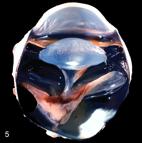
ネコ新生血管硝子体網膜症の病理画像。網膜剥離と出血を示唆する色素が網膜上に見られる(Figure 3より)
コメント
教科書を除いて初めての査読あり論文でのネコ新生血管硝子体網膜症(Feline neovascular vitreoretinopathy:FNV) の報告であり、そのケースシリーズです。
0
